

How a FIB-SEM works

I’m going to explain how a FIB (focused ion beam) - SEM (scanning electron microscope) works by posting screenshots of my conversation with my 13yo kid, explaining how it works.
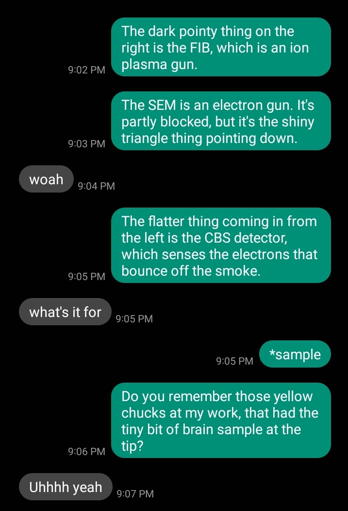
This was the picture I sent:

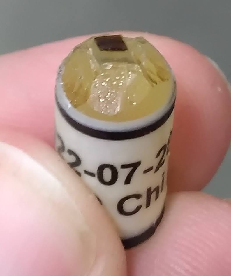
I’m referring to one of these:
So, to recap, we have:
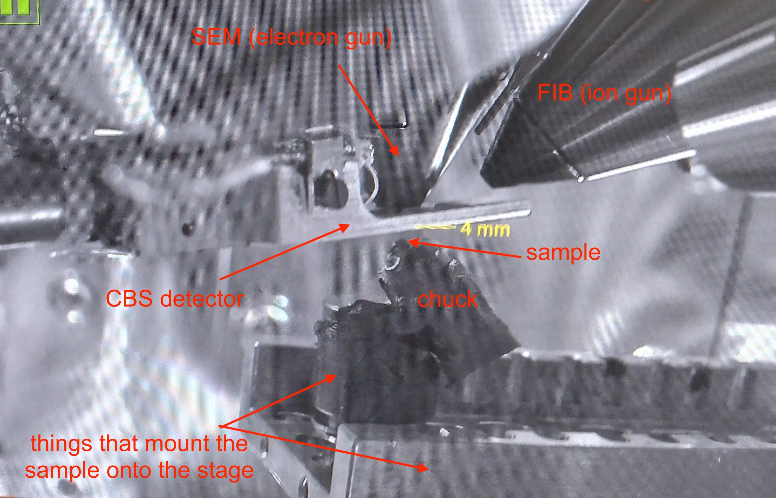
Now that I’ve told her what all the parts are, I start the explanation of how they work.

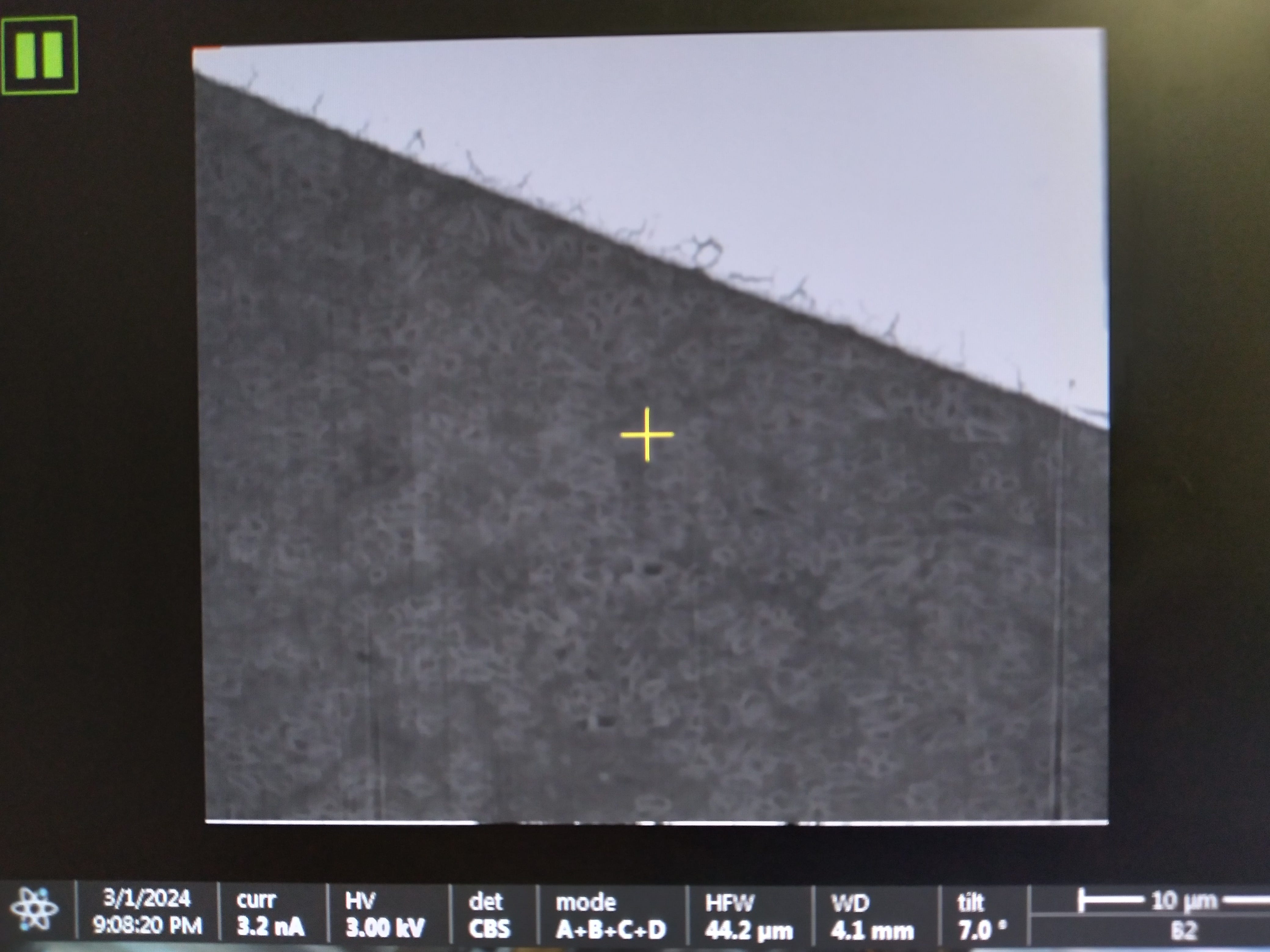
And then I sent this pic:

Since I didn’t get around to responding to that first question, let me do it now. When you shoot electrons at an insulator, they have nowhere to go, and soon you end up with a giant cloud of electrons pushing all your new electrons back into the detector, making pictures where all you can see is a giant spasm of white.
This is called “charging”, and one of the ways to stop it is to coat your sample in something conductive, so that incoming electrons can flow away into the universe. Carbon-coating is more common, but for this sample, my FIB-SEM teacher taught me how to use the gold-coater.
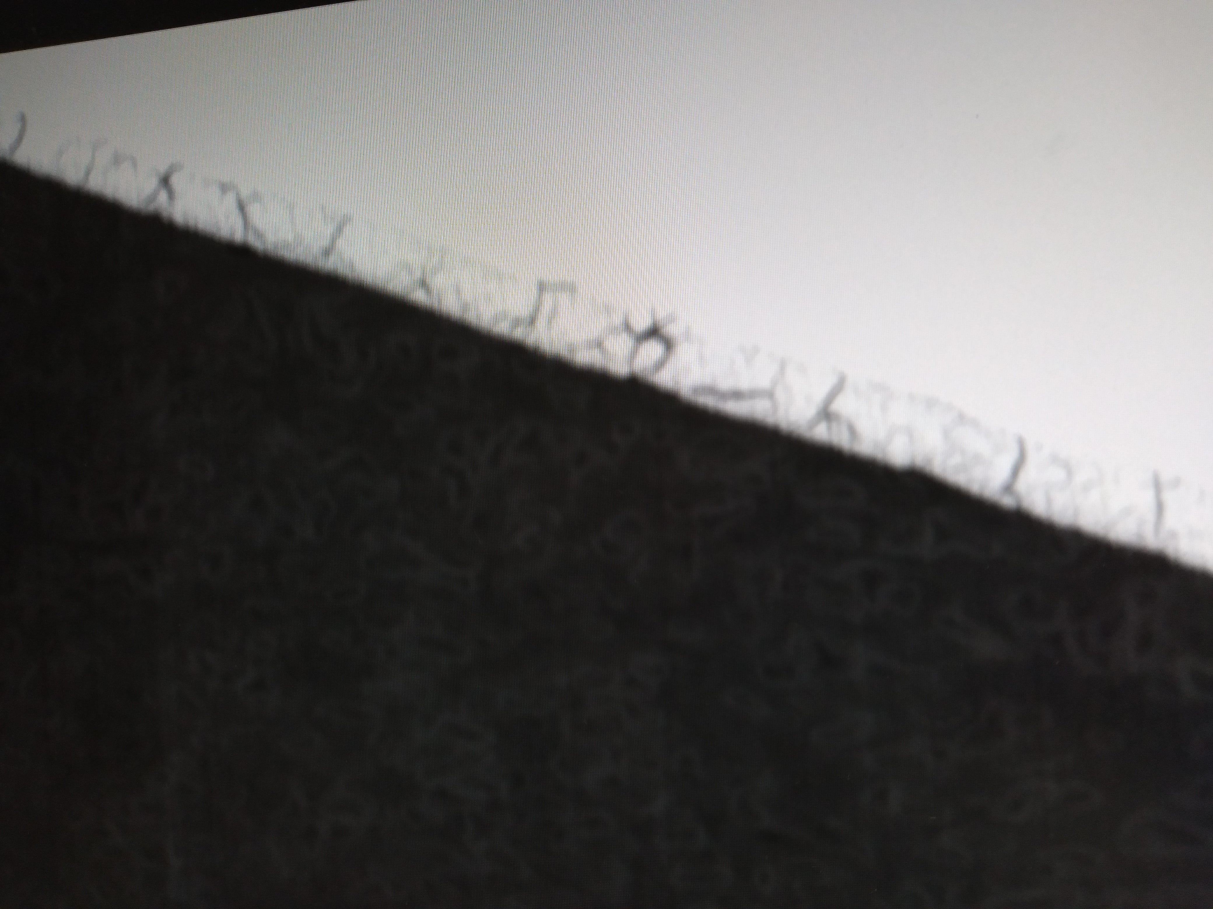
Her second question, about the hairy things, I do go on to answer.

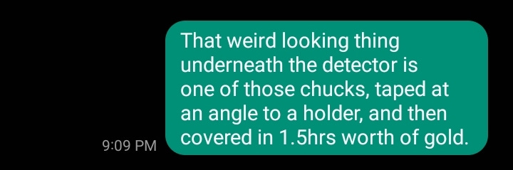
And then I sent this picture:
My kid’s response:

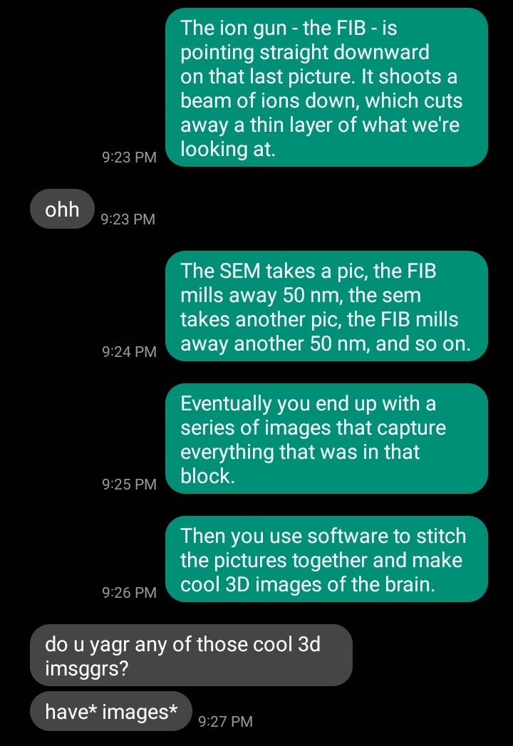
I didn’t, at the time, but now I do. Here’s a couple examples of the kinds of images you can get.